New Methods of Diastatic Detection
Cover your buts: Fix alarming gaps in existing testing and get more helpful info

Jul 28, 2025

Three New Tests
Say you’re screening via LCSM, which, with proper validation and optimization, is dialed in to catch as many diastatic strains as possible. You get some growth — a positive hit. Now what?
Pick up that colony, and apply one of these relatively easy-to-incorporate new tests, which will help you both resolve false results and see how diastatic a detected contamination is:
Monitor pH drop in a liquid solution containing maltodextrin and Bromocresol green dye indicator.
Measure how much dextrin is consumed over time in a liquid solution using iodine staining (and UV-vis Spectrophotometry).
Watch for growth, including cross-feeding using the non-diastatic indicator strain W303 on a plate culture.
Monitor pH Drop with Bromocresol Green Dye
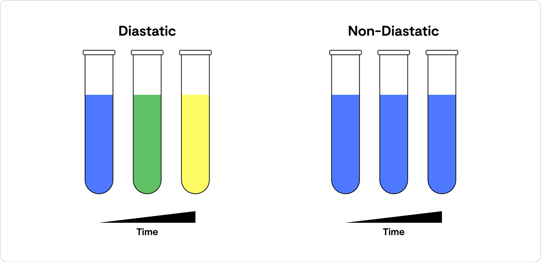
Developed in collaboration with Omega Yeast’s friends at Cornell, this method is based on diastatic yeast growth in the presence of the pH indicator dye Bromocresol green.
If the inoculating yeast is diastatic, it successfully breaks down the dextrin supply into digestible glucose and begins to grow and replicate. That causes a pH drop. The pH changes the color of the solution from blue to green to yellow. The color change confirms it’s diastatic; how fast and fully it changes tells you how aggressively diastatic.
Note: to avoid the source of simple sugars that plate agar naturally adds, Omega Yeast’s R&D team tried a non-starch, liquid medium. They used maltodextrin as the complex sugar supply. Though any dextrin supply is likely impure enough to have some easily fermentable sugars, whatever impurity is present doesn’t support enough growth to confuse this test.
In Experiments
At a starting pH of around five or six, the bromocresol green dye is blue. Inoculating the test medium with a non-daistatic control strain resulted in the pH indicator’s color remaining blue. With a strongly diastatic strain like French Saison, however, color-change happened very quickly.
How quickly strains’ growth affected the pH of the solution varied, showing the differences in the speed with which diastatic strains break apart maltodextrin chains into fermentable sugars to use as fuel for reproduction.
Diastatic strains were considered ‘strongly’ diastatic when the solution turned completely yellow within 3 days, ‘moderately’ diastatic when it turned yellow within 8 days and ‘weakly’ diastatic when a color change occurred, but was incomplete even after 2 weeks.
Measure Dextrin with Iodine Staining and UV-vis Spectrophotometry
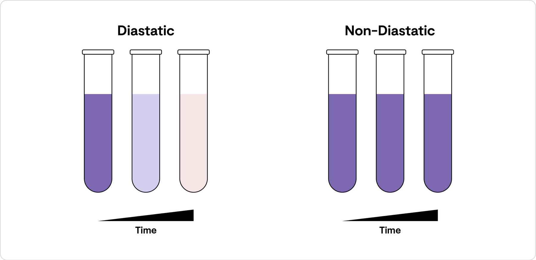
This method uses dextrin as the sole food source in a liquid solution, to which Omega Yeast’s researchers added iodine stain, an indicator dye that turns starches purple. The more whole dextrin there is, the darker the purple-color is. As dextrin is broken up by the enzymatic activity of diastatic yeast, the iodine’s purple color becomes lighter. If there’s no color change, then there’s no diastatic activity. The speed of the color change is indicative of diastatic intensity.
With UV-vis spectrophotometry, you can measure how much dextrin is remaining according to the saturation of the dye indicator color.
In Experiments
Diastatic strains were considered ‘strongly’ diastatic when more than 50% of the dextrin present was broken down within a couple of weeks. They were classified as ‘moderately’ diastatic when that took around three weeks, and ‘weakly’ diastatic when dextrin degradation hadn’t reached 50% by week 4.
Cross-Feeding to a Non-Diastatic Lab Indicator Strain
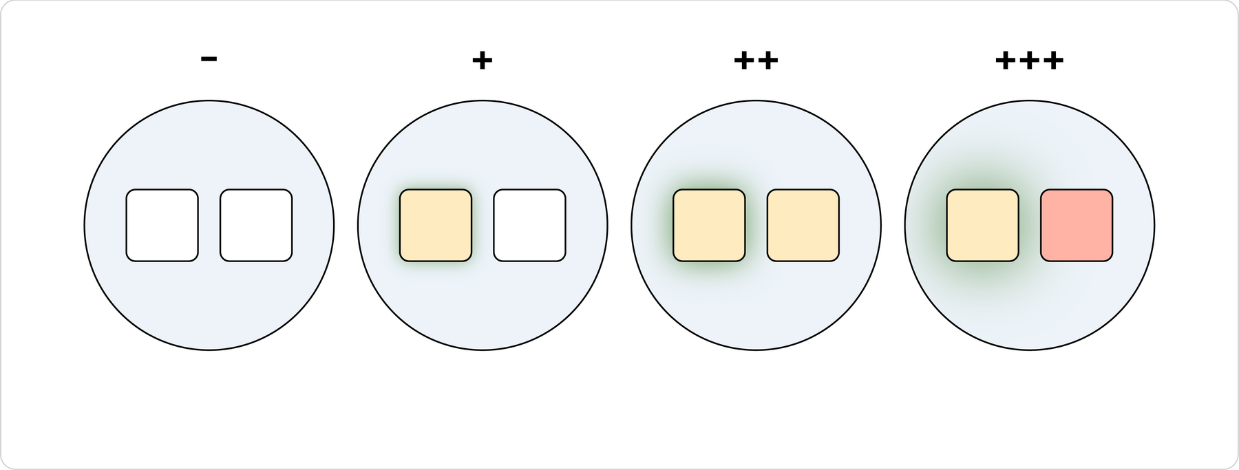
This test shows whether a strain is diastatic or not by its successful growth on a plate where dextrin is the main food source. When the test strain is patched next to a non-diastatic indicator strain, the test strain feeds the non-diastatic strain the fermentable sugars it sources from dextrin.
The combination detects both the presence of diastatic yeast, and its diastatic intensity, because the non-diastatic indicator strain, a laboratory workhorse called W303, happens to accumulate red color as it grows. When it has grown robustly, that growth is visible thanks to a red color appearing.
Neither the test strain, nor the passenger indicator strain would grow unless the test strain was diastatic.
In Experiments
- With a strongly diastatic strain, both strains grow. The glucoamylase enzyme dilutes into the medium and breaks dextrins down into glucose, feeding both the diastatic strain and the indicator strain. The indicator strain turns red thanks to strong growth.
- With a moderately diastatic strain, cross-feeding begins to occur, but not to the same degree. The diastatic strain grows, and the indicator strain grows, but may or may not redden.
- A weakly diastatic strain may secrete enough enzyme to allow for its own growth, but not enough to spur the growth of the indicator strain.
- With a non-diastatic strain, neither it nor the indicator strain really grow (other than what little they can scrounge from unintentionally fermentable plate sugars).
| Description | Method 01 | Method 02 | Method 03 | Bottle Refermentation |
|---|---|---|---|---|
| British Ale I | - | nd | nd | nd |
| Neutral Ale | - | - | - | - |
| French Saison | +++ | +++ | +++ | +++ |
| Jovaru | ++ | ++ | ++ | ++ |
| Biere de Garde | +++ | ++ | ++ | ++ |
| Belgian Strong Ale | +++ | +++ | ++ | +++ |
| Swiss Ale | +++ | +++ | ++ | +++ |
| Specter Ale | +++ | +++ | ++ | ++ |
| Saisonstein | +++ | +++ | +++ | ++ |
| Gulo | +++ | +++ | ++ | - |
| Belgian Ale D | ++ | + | + | ++ |
| Bavarian Wheat | ++ | ++ | + | ++ |
| Belgian Saison I | +++ | ++ | ++ | + |
| Belgian Dark Ale | + | - | - | + |
| Belgian Saison II | +++ | ++ | ++ | ++ |
| Vermont Farmhouse | +++ | ++ | ++ | + |
| Tropical IPA | ++ | + | + | ++ |
Related Collections

